Fig. 7.
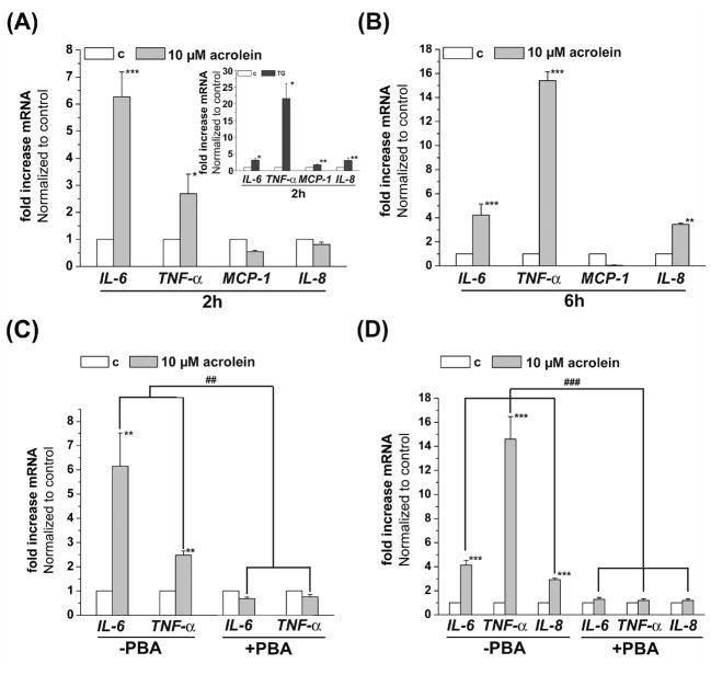
Acrolein increases the transcription of inflammatory cytokines and chemokines by endothelial cells via ER stress. HUVECs were treated with 10 μM acrolein and harvested after 2 (A) or 6 (B) h as described in Materials and Methods. Levels of IL-6, IL-8, TNF-α and MCP-1 mRNA was quantified by RT-PCR of 3 (2h) or 6 (6h) independent experiments. Thapsigargin (1 μM, 2h) treated HUVECs were used as positive controls (A, insert) for the 2h treatment. Inhibition of the acrolein induced cytokine and chemokine transcription by PBA. HUVECs were left untreated or pretreated with 10 mM PBA and then incubated with 10 μM acrolein. Two (C) or 6 (D) hours after acrolein treatment, the cells were harvested and levels of IL-6 and TNF-α mRNA (2h) or IL-6, IL-8 and TNF-α-mRNA (6h) were examined by RT-PCR. Data from 3 to 5 independent experiments were analyzed and presented as values normalized to control. Data are presented as mean ± SE, * p<0.05, ** p<0.01, ***p<0.001, and ### p<0.01, ### p<0.001 PBA treated versus untreated cells.

