Fig. 2.
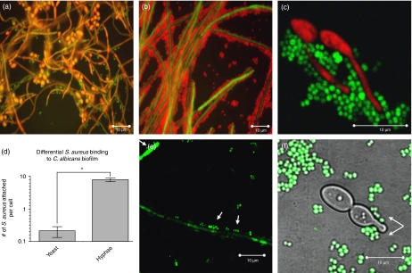
Biofilm architecture of Candida albicans and Staphylococcus aureus 24-h dual-species biofilm using PNA-FISH and GFP-expressing microorganisms. (a) Staphylococcus aureus (FITC-labeled probe, green) has a greater tropism for the hyphal form of C. albicans (TAMRA-labeled probe, red) compared with the yeast form. Field of view diameter is 150 μm. (b) An area of C. albicans (FITC-labeled probe, green) hyphal biofilm growth is completely covered by S. aureus (Cy3-labeled probe, red). (c) A × 63 zoom image showing staphylococci (FITC-labeled probe, green) binding to only the hyphal filaments of C. albicans (Cy3-conjugated probe, red). (d) Graph representing the average number of S. aureus cells attached per C. albicans cell during polymicrobial biofilm growth. Ten fields were chosen at random for counting and the experiment was repeated in triplicate. Error bars represent the SD. (e) Staphylococcus aureus (white arrows), expressing GFP under control of the sarA promoter, was found to be associated to GFP-expressing C. albicans hyphae. (f) Staphylococcus aureus (white arrows) demonstrating preferential binding to a C. albicans germ tube without binding to the yeast cell. Fluorescence was captured with a × 63 oil-immersion objective and FITC/DICIII, FITC/Texas Red filter sets. Asterisk (*) denotes a statistically significant difference at P<0.05.

