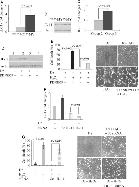
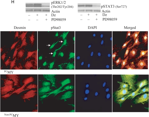
Figure 1.
IL-11 expression in PCMY and its role in survival signalling. (A) Real-time PCR showing 3.72-fold higher expression of IL-11 in vitro in PCMY (P = 0.013 vs. non-PCMY; n = 3) which was confirmed by (B) western blot studies. (C) Real-time PCR showing 1.91-fold up-regulation of IL-11 in the LV of Group 3 animal hearts when compared with Group 2 (P = 0.004; n = 3/group) 4 days after their respective treatments. (D) Western blotting revealed up-regulation of IL-11 protein in PCMY which was sensitive to Erk1/2 inhibition by PD098059. Pre-treatment of the cells with PD98059 for 30 min abrogated IL-11 expression with resultant loss of cytoprotection against oxidant stress. (E) Treatment of MY with Dz alone had no cytotoxicity on the cells; however, significantly higher LDH release was observed in non-PCMY upon treatment with H2O2 when compared with PCMY (P = 0.004; n = 5). Pre-treatment of PCMY with PD98059 significantly diminished the cytoprotective effects as well as morphological integrity of the cells. (F) RT–PCR showing successful knock-down of IL-11 expression in MY by IL-11-specific siRNA transfection Dz preconditioning of the cells failed to induce IL-11 expression. Sc siRNA-transfected cells were used as a control. (G) Treatment with H2O2 caused significantly more LDH leakage from IL-11-silenced cells when compared with its preconditioned counterpart (P = 0.013; n = 3) with concomitant morphological distortions. (H) Western blot analysis showed that reconditioning activated Erk1/2 and Stat3, which was evident from higher level of phosphorylation. Pre-treatment of cells with PD98059 abolished their phosphorylation upon subsequent treatment with Dz. Treatment with H2O2 caused significantly more LDH leakage from cells pre-treated with PD98059 when compared with its preconditioned counterpart (P = 0.004). Fluorescent immunostaining for pStat3 expression revealed that preconditioning promoted pStat3 (green fluorescence; indicated by white arrows) localization in the nucleus. The cells were stained for desmin expression (red fluorescence) to show the purity of skeletal myoblasts. The nuclei were visualized by DAPI staining (blue fluorescence). In non-PCMY, pStat3 was missing in the nucleus.


