Figure 3.
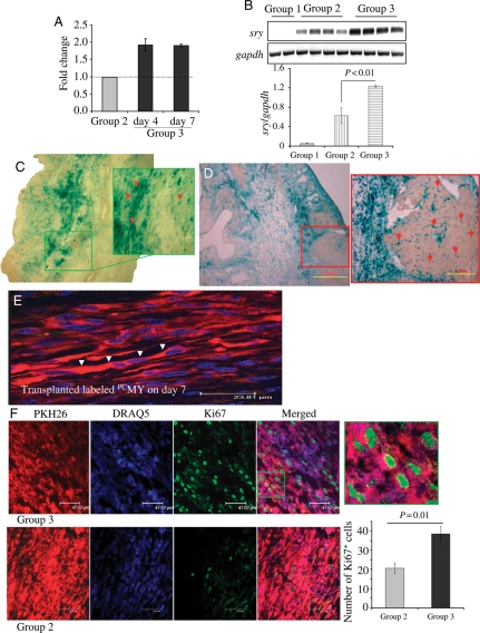
Enhanced survival and proliferation of PCMY in the infarcted heart. Quantification of cell survival by (A) real-time PCR and (B) conventional PCR for donor-specific sry-gene. A significantly higher survival of PCMY (Group 3) was observed on 4 and 7 days post-transplantation, respectively (n = 4 on each time point) when compared with non-PCMY (Group 2; n = 4 on each time point; P < 0.01 vs. non-PCMY). DMEM without cells injected Group 3 (n = 2 on each time point) did not show sry-gene expression and served as a negative control. (C and D) Tracking of nlsLacz-labelled transplanted cells by β-gal histochemistry at 7 days post-transplantation (C) non-PCMY transplanted and (D) PCMY transplanted tissue sections (original magnification, ×100). Red and green boxes in the respective figures represent the magnified regions with red arrows showing nlsLacz-labelled transplanted cells. (E) Histological tissue section from Group 3 animal heart 7 days after PCMY transplantation showing multinucleated myotubes formed from PKH26-labelled transplanted cells (red). The nuclei were stained blue with DAPI (original magnification, ×1000). (F) Preconditioning increased the proliferative capacity of transplanted cells. Fluorescence immunostaining for Ki67 antigen on heart tissue sections from 7 days post-transplanted animals (red, PKH26-labelled cells; green, Ki67-positive cells; blue, DAPI for nuclear staining).

