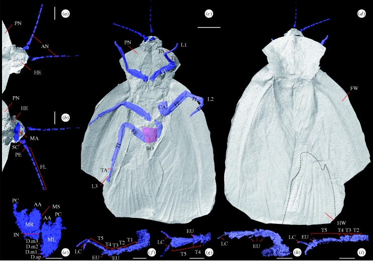
Figure 2.
Computer reconstructions of A. eggintoni. (a) Dorsal view of anterior. (b) Ventral view of anterior. (c) Ventral view, showing all limbs and body. (d) Dorsal view showing wings, pronotum and head. (e) Mandibles. Looking posteriorly, labelling after Zhuzhikov (2007). (f) Right foreleg in the lateral view. (g) Right ventral foreleg. (h) Right midleg in the lateral view. (i) Right ventral midleg. Scale bars, (a,b) 1 mm; (c,d) 5 mm, (e–i) 0.5 mm. All images from the higher resolution model except (c,d). AA, anterior acetabulum; AN, antennae; CA, coxa; D.ap, apical tooth; D.m1–D.m3, marginal teeth; EU, euplantulae; FE, femur; FL, flagellum; FW, forewing; HE, head; HW, hindwing; IN, incisor; L1, foreleg; L2, midleg; L3, hindleg; LC, lateral claw; MA, mandibles; ML, left mandible; MR, right mandible; MS, molar surface; PC, posterior condyle; PE, pedicel; PN, pronotum; SC, scape; T1–5, tarsomeres 1–5; TA, tarsus; TI, tibia.

