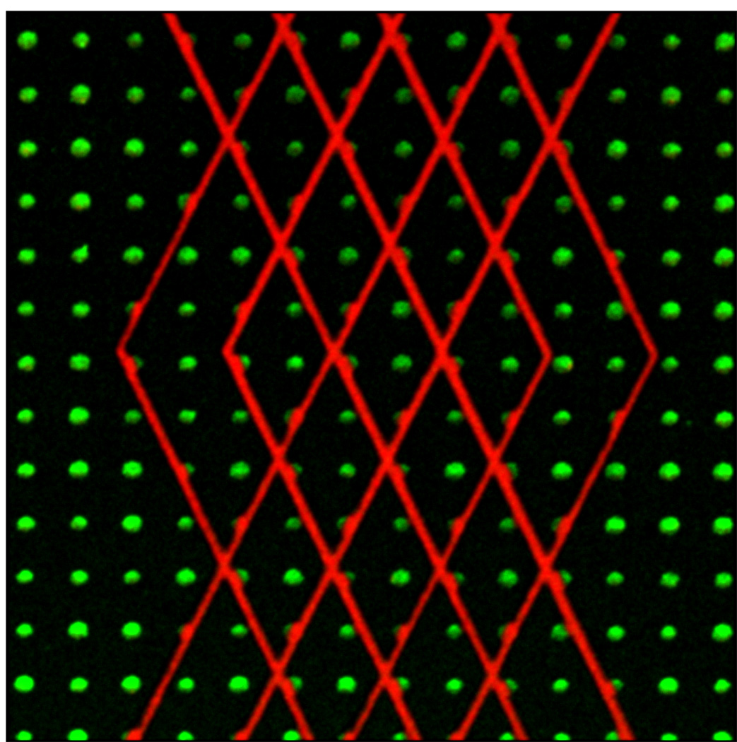
Figure 23.
Dual ECM patterns by performing µPP twice in series. A first ablation was performed followed by quenching, ECM adsorption, and blocking. A second round of ablation was done is the presence of the second ECM. Green dots are fibrinogen and red lines are vitronectin. Dots are spaced 5 microns apart.

