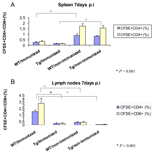
Figure 7.
Flow cytometric analysis of recipient mouse spleens and lymph nodes. A) The percentage of CD4+ and CD8+ T cells in the spleens of mice receiving immunized and non- immunized donor cells. B) The percentage of CD4+ and CD8+ T cells in the lymph nodes of the recipient mice. The cells were surface stained with anti-CD3+ and anti-CD4+ antibodies or anti-CD3+ and anti-CD8+ and analyzed by flow cytometry (P <0.001).

