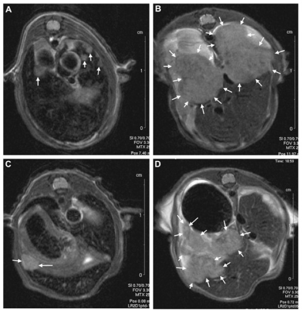
Figure 5.
Viral replication in the liver tumors. The growth of liver metastasis was analyzed with magnetic resonance imaging (MRI). Tumors are marked with arrows. Picture of liver metastasis of mock treated (PBS) animal (A) 1 day before treatment and (B) on day 35 after treatment. (C) Picture of liver metastasis of WT-RGD treated animal one day before treatment and (D) on day 35 after WT-RGD treatment.

