
Figure 2.
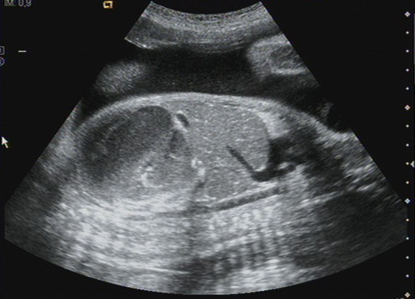
Ultrasound image: 32.3 weeks of gestation. Longitudinal scan image thorough the fetal abdomen identifying a mass occupying the entire left hemiabdomen (meconium pseudocyst), with mixed echogenicity. No calcifications were observed.
Official websites use .gov
A
.gov website belongs to an official
government organization in the United States.
Secure .gov websites use HTTPS
A lock (
) or https:// means you've safely
connected to the .gov website. Share sensitive
information only on official, secure websites.
Ultrasound image: 32.3 weeks of gestation. Longitudinal scan image thorough the fetal abdomen identifying a mass occupying the entire left hemiabdomen (meconium pseudocyst), with mixed echogenicity. No calcifications were observed.