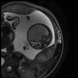
Figure 4.

Magnetic resonance imaging: 32.4 weeks of gestation. Transverse magnetic resonance image of the fetus demonstrating the meconium pseudocyst; a 72 × 58 mm, heterogeneous, mesenteric mass without necrosis causing significant distortion of the small intestine to the left. No calcification or ascites were observed.
