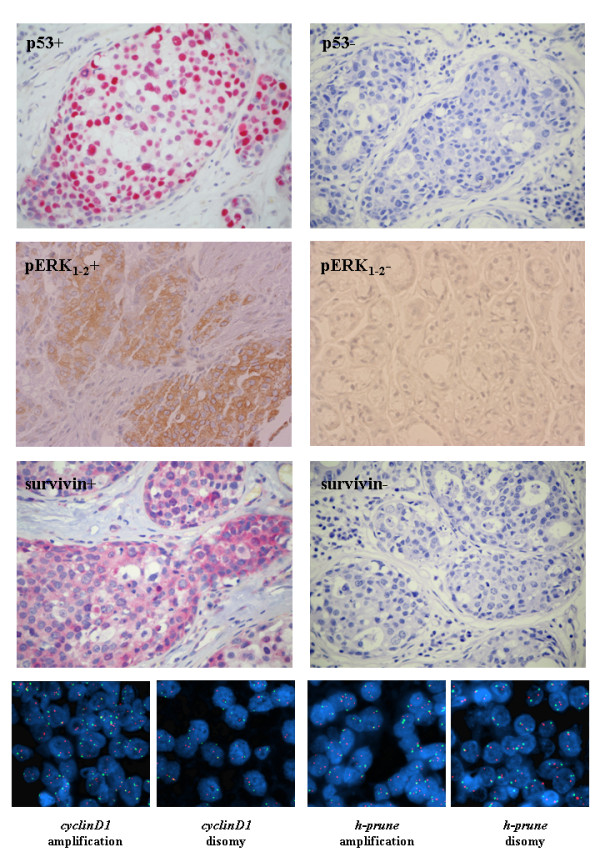
Figure 1.

Immunohistochemistry and FISH analysis. (Up-middle) Typical examples of T4 breast carcinoma tissue sections positive (left) or negative (right) for p53, pERK1-2, and survivin protein expression. (Bottom) Typical examples of double-colour FISH results. Nuclei extracted from paraffin-embedded tissues after hybridization with probes specific for cyclinD1 or h-prune loci (red signals) and control chromosome centromeres (green signals).
