Figure 2.
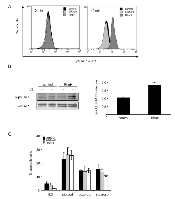
RhoH expression triggers IL3-dependent STAT1 activation. (A) Control cells, siRhoH and RhoH-transduced BaF3 cells were depleted of cytokine and FBS for 3 h prior to stimulation with IL3. Phosphorylated STAT1 levels were measured with intracellular FACS staining using a FITC-labelled pTyr-STAT1 specific antibody. One representative result of three independent experiments is shown. (B) Control and RhoH cells were starved for 3 hours prior to stimulation with 50 ng/ml IL3. Cells were lysed, STAT1 was immunoprecipitated and tyrosine phosphorylated STAT1 and total STAT1 were detected by enhanced chemiluminescence. Quantification of pSTAT1 levels was done after normalisation to STAT1 levels in cells transduced with the empty vector and is presented as induction of phosphorylation compared to the corresponding unstimulated sample. Statistical significance was analysed using the 's t-test (mean ± SD, n = 3; ***P ≤ 0.005). (D) Apoptosis was induced in control cells, siRhoH or RhoH-transduced cells by cultivation in the absence of IL3 (overnight), treatment with 1 μM staurosporine (3 h) or with 1 μM doxorubicine (12 h) prior to Annexin V/PI staining and FACS analysis (mean ± SD, n = 3).

