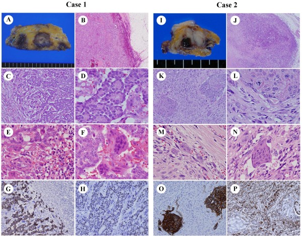
Figure 1.
Gross and microscopic findings of breast carcinoma with OGCs. OGCs in Case 1 appeared associated with invasive ductal carcinoma, grade 1, and in Case 2 with carcinosarcoma. A-H: Case 1. A: Gross appearance. B: Low power view of the tumor. C, D: High power views of the tumor. E, F: Bi-, tri-, tetranucleate cells were observed as well as OGCs in hypervascular inflammatory stroma. G: CK AE1/AE3 staining. H: ER staining. I-N: Case 2, corresponding to A-F in Case 1. O: CK AE1/AE3 staining. P: Vimentin staining.

