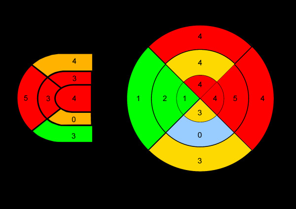
Figure 1.
Distribution of histological inflammation. Bullseye plots showing distribution of histologically verified inflammation inside right (A) and left (B) ventricular wall from apex (internal segment) to heart base (external segments). Myocardial inflammation was predominantly located in the anterior and lateral wall of right and left ventricle.

