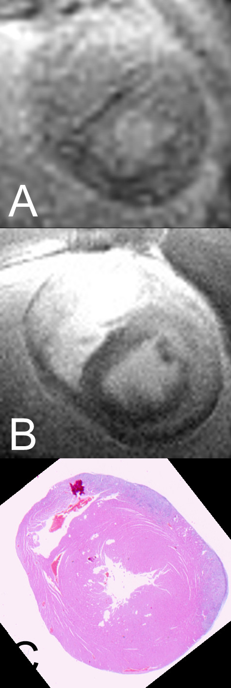
Figure 3.

Sample images of CMR. CMR images and histopathological finding of an animal from the experimental group: (A) Turbo-FLASH-examination without fat saturation. (B) TSE-examination without fat saturation. (C) Histopathological findings with visible infiltration in the left anterior.
