Figure 2.
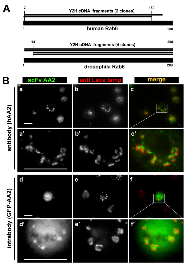
Characterization of the scFv AA2 (anti-Rab6•GTP). A. Schematic alignment of human and drosophila Rab6 with translated sequences of their respective cDNA of the interacting clones (numbers indicate amino acid position, vertical lines project the SID onto the full length sequences). Note that in both cases the SID spans the entire core of the respective open reading frames (encoding for a protein of 208 amino acids each). B. AA2 recognizes the drosophila Rab6 homologue as antibody and as intrabody. AA2 as antibody: S2 insect cells were fixed, stained with hAA2 (a, green in c) and co-stained with anti-Lava lamp (b, red in c). Panels a', b' and c' represent a magnification of an area within a, b and c. Rab6-containing Golgi membranes are decorated with hAA2 and surrounded by Lava-lamp-positive structures. AA2 as intrabody: S2 cells were transfected with a plasmid expressing AA2 fused with GFP (d, green in f). Eighteen hours later cells were fixed and co-stained with anti-Lava lamp antibodies (e, red in f). Panels d', e' and f' represent a magnification of an area within d, e and f respectively. AA2 is a functional intrabody in insect cells as it stains Golgi membranes like the AA2 antibody. Stronger labeling of Golgi structures with GFP was seen in living cells (data not shown). Bar 10 μm.

