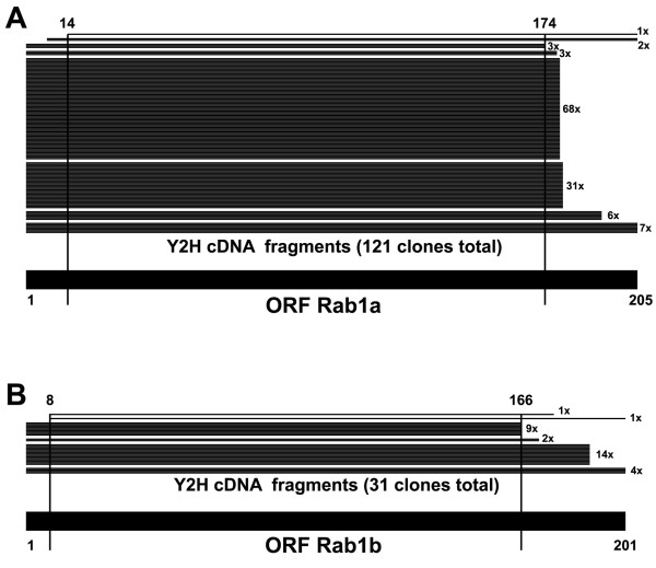
Figure 3.
Characterization of the scFv ROF7 (anti-Rab1•GTP). Schematic alignment of human Rab1a (A) and Rab1b (B) with the translated sequences of cDNA fragments found in the ROF7 screen (numbers indicate amino acid position, vertical lines project the SID onto the full length sequences). Note that in both cases the SID spans the entire core of the respective open reading frames (encoding for proteins of 205 and 201 amino acids, respectively).

