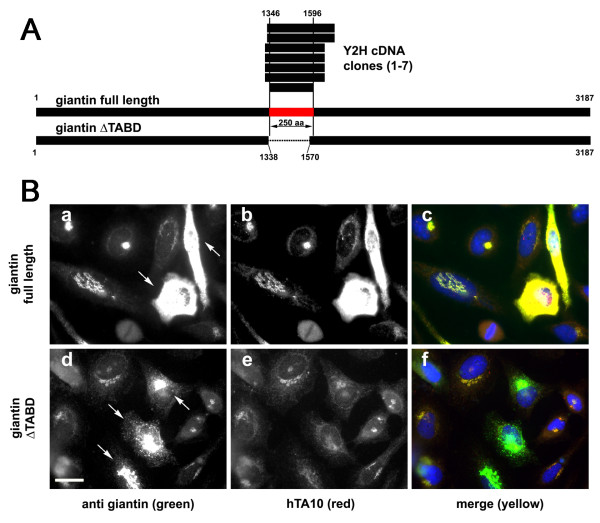
Figure 4.
Characterization of scFv TA10 (anti-giantin). A. Schematic alignment of full-length rat giantin with the translated sequence of the seven clones obtained from the TA10 screen (above) and the deletion construct used to confirm the localization of the interaction domain (below). Numbers indicate amino acid position relative to the full-length rat giantin sequence; horizontal lines project the SID (~28 kDa) onto the giantin sequence. This 250 amino-acid-long stretch is depicted in red. B. Confirmation of the ~28kDa large SID as the region which contains the TA10 epitope. HeLa cells were transfected either with full-length rat giantin (a-c) or with a giantin construct lacking identified SID (d-f). Cells were fixed 18 hours after transfection and double stained using a commercial anti-giantin antibody (green) and with hTA10 (red). Overexpression of the full-length protein was detected by hTA10 (b and c), while the staining was unchanged or even diminished in cells transfected giantin ΔTABD (e and f). Arrows indicated cells that strongly overexpress the respective constructs. Bar 10 μm.

