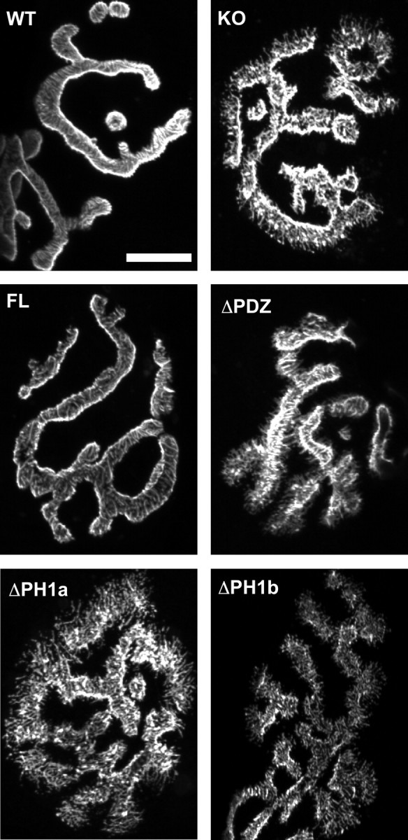
Figure 2.

Transgenic rescue of aberrant NMJ structure in the α-syntrophin KO muscle. En face views of NMJs from the sternomastoid muscle visualized using fluorescent α-bungarotoxin. The spikes observed in the α-syntrophin KO mice are not present in the FL transgenic, which has continuous smooth edges similar to those of the WT mouse. NMJs of mice lacking the PDZ or either portion of the PH1 domain resemble the KO with spicules and fragmented edges. Scale bar, 20 μm.
