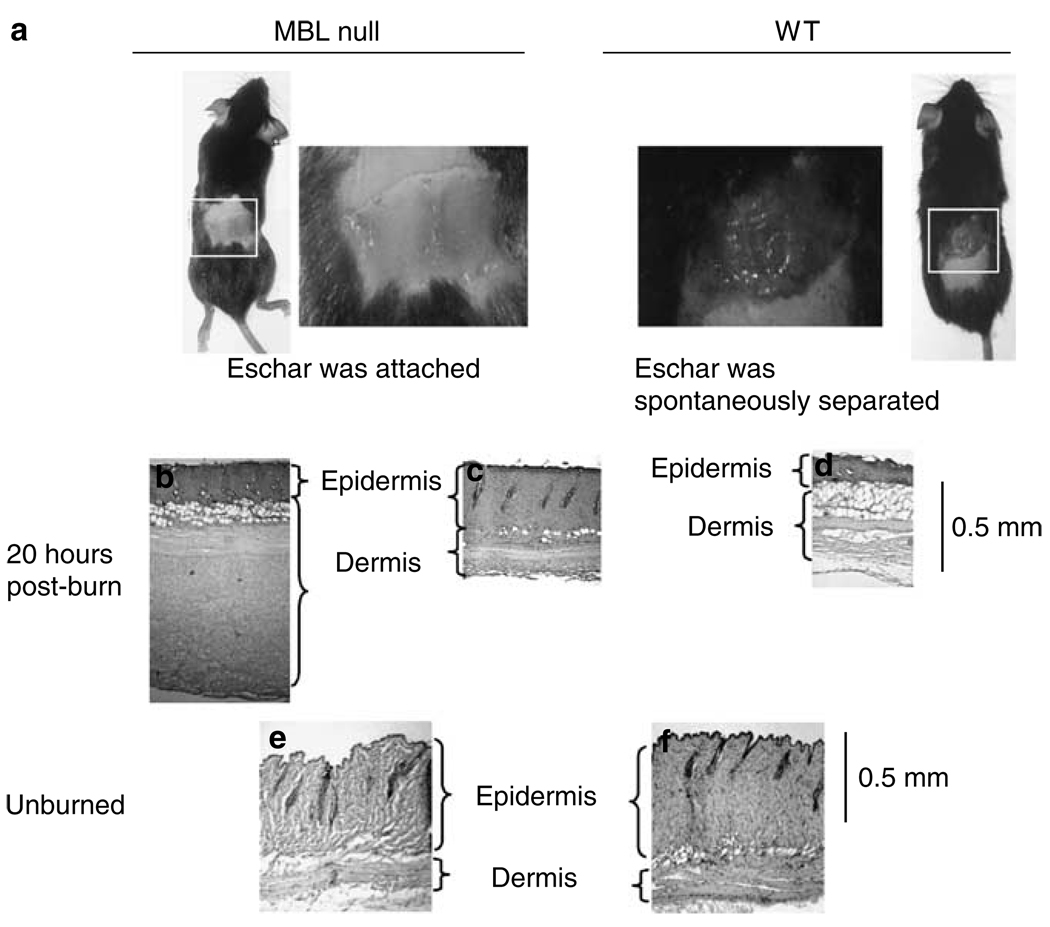
Figure 1. Eschars in WT and MBL null mice after burn.
(a) Phenotype of MBL null mice and WT mice 20 hours postburn. Histological examination of eschars after 20 hours by hematoxylin and eosin staining; (b) and (c) MBL null mice; (d) WT mice. Normal skin without burn; (e) MBL null mice; and (f) WT mice.

