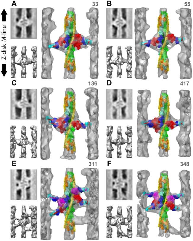
Figure 3. Diversity of myosin-actin attachments shown in quasiatomic models of reassembled primary class averages.
The number in the upper right is the number of the corresponding raw repeat, NOT the number of raw repeats averaged within the class. Small panels to the left are the central section and an opaque isodensity surface view of the larger panel without the quasiatomic model. Actin long pitch strands are cyan and green with the target-zone actins in darker shades, TM is yellow and Tn orange. Strongly bound myosin heads are red, weak binding myosin heads are magenta, The essential light chain is dark blue and the regulatory light chain light blue. (A) shows a single headed cross-bridge on the left and a 2-headed, strong binding cross-bridge on the right. (B) shows a pair of 1-headed, strong-binding cross-bridges on actin subunits H and I. (C & D) have a 2-headed cross-bridge on the left and a 1-headed cross-bridge on the right, all strongly bound to actin. (E & F) are mask motifs with Tn-bridges. In (E) the right side M-ward weak binding cross-bridge is bound outside of the target zone to TM near actin subunit F while the one on the left is within the target zone on actin subunit I. In (F), the weak binding, left-side, M-ward cross-bridge is bound outside the target zone to TM near actin subunit G while the weak binding cross-bridge on the right is bound to target-zone actin subunit H. Tn-bridges have not been fit with a myosin head. These six reassembled repeats can also be viewed in Supporting Movies S1, S2, S3, S4, S5 and S6.

