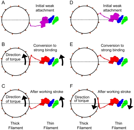
Figure 10. Two mechanisms to account for azimuthal skewing of lever arms of strongly bound cross-bridges.
(A–C) Conversion from weak-to-strong binding according to Scenario 1. (D–F) Active azimuthal component to the working stroke. View direction is M-line toward Z-line. Myosin is colored either red (strong binding) or magenta (weak binding). Actin subunits are green and blue. Three successive levels of S2 origins are shown in shades of brown that darken with distance from the observer. The lever arm is the line originating on the red (or magenta) MD while S2 is shown as a short segment when oriented nearly parallel with the filament axis and becomes longer when angled with respect to the filament axis. The horizontal line is the inter-filament axis. Arrows show the direction of the torques (not their magnitude) produced during the weak-to-strong transition or as a component of force generation and filament sliding. The direction of thin filament movement during sarcomere shortening is toward the observer. (A & D) Initial weak binding is shown, which in (A) begins away from the strong binding orientation and in (D) begins in the strong binding orientation but with actin binding cleft open. (B) Conversion to strong binding involves diffusion of the MD clockwise on actin which swings the S2 anticlockwise about the thick filament. (C) Force production realigns S2 with the filament axis while bending the lever arm azimuthally. (E) Transition from weak to strong binding involves no change in myosin orientation on actin, just a closing of the actin binding cleft. The lever arm in (D & E) is in the same orientation suggested by the crystal structures. (F) An azimuthal component to the working stroke moves the lever arm clockwise around the thick filament. This figure can be seen as an animated sequence in Supporting File S1.

