Fig. 2.
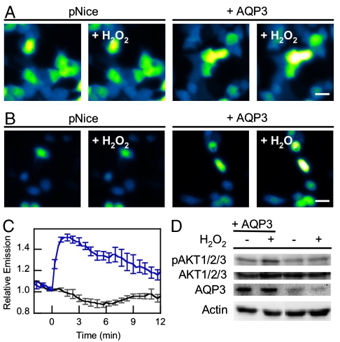
AQP3 mediated exogenous H2O2 uptake can effect intracellular signaling. (A) Live-cell imaging of changes in HyPer fluorescence upon treatment of HEK 293 cells with 10 μM H2O2. “pNice,” HEK 293 cells expressing HyPer and transfected with pNice as a control vector before addition of H2O2. “+AQP3,” HEK 293 cells expressing HyPer and AQP3 before addition of H2O2. “+H2O2,” cells 2 min after addition of 10 μM H2O2. 20 μm scale bar. (B) Live-cell imaging of changes in HyPer fluorescence upon treatment of HeLa cells with 10 μM H2O2. “pNice,” HeLa cells expressing HyPer and transfected with pNice as a control vector before addition of H2O2. “+AQP3,” HeLa cells expressing HyPer and AQP3 before addition of H2O2. “+H2O2,” cells 2 min after addition of 10 μM H2O2. 20 μm scale bar. (C) Time-course and quantification of (B). HeLa cells expressing HyPer and transfected with pNice (black line) or AQP3 (blue line) were stimulated with 10 μM H2O2 at t = 0 and the changes in HyPer fluorescence monitored over time. Error bars are ± s.e.m. (n = 3). (D) Western blot showing changes in pAKT1/2/3 levels in HeLa cells treated with H2O2. HeLa cells expressing AQP3 or control vector were serum-starved and then treated with 200 μM H2O2 for 20 min at 37 °C and then lysed. Phospho-AKT and AQP3 levels were measured by Western blot analysis of whole cell extracts, followed by stripping and reprobing for total AKT or Actin, respectively.

