Fig. 2.
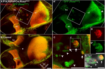
Activation of Notch leads to ectopic patches of hair cells in nonsensory regions. Ectopic clusters of MyosinVIIa-labeled hair cells form in nonsensory regions of the vestibular epithelia of hGFAPCre;RosaNotch mice. Confocal projection views of vestibular epithelia from P14 hGFAPCre;RosaNotch (A and A’) or hGFAPCre/+ control (B) mice, including the utricle (ut), horizontal crista (hc), and anterior crista (ac) immunolabeled with anti-GFP (green) and the hair cell-specific marker anti-MyosinVIIa (red). In the hGFAPCre;RosaNotch epithelium, regions of ectopic hair cell formation are present between the utricle and cristae. In control vestibule, the same region (B, asterisk) is always nonsensory and devoid of hair cells. (C) High-magnification view of the boxed region in A, which contains multiple clusters of GFP+ cells (arrowheads mark examples), three of which contain ectopic MyosinVIIa+ hair cells (arrows and box). (D and D’) Split-channel views of the boxed region in C. (E) Tuj1 immunolabeling (white) shows vestibular neurites projecting to the ectopic patches in the upper region of C, as shown as a z series in Movie S2. [Scale bars, 100 μm (B, C, and E) and 50 μm (D’).]

