Fig. 2.
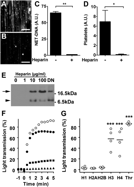
Heparin dismantles NETs and prevents histone induced platelet aggregation. SytoxGreen staining of NETs perfused for 10 min with blood in the absence (A) or presence (B) of heparin. (Scale bars, 100 μm.) (C) Quantification of NETs after 10 min perfusion with normal (-) or heparinized (+) blood. (D) Quantification of platelets on NETs perfused for 10 min with blood before (-) and after (+) treatment with heparin. Data presented as mean ± SEM, n = 3; (Student's t test; *P < 0.05; **P < 0.01). (E) Heparin and DNase released histones from NETs. Immunodetection of histone H2B (arrow) in the culture supernatants of NETs treated with heparin or DNase (DN). A second band (arrowhead) may represent cross reactivity of the antibody or a proteolytic product. Data presented are representative of three independent experiments. (F) Aggregometry of platelets stimulated with thrombin (open circles) or human recombinant histone H3 (solid circles). EDTA (solid squares) and heparin (solid triangles) inhibited platelet aggregation by histone H3. (G) Extent of platelet aggregation 3 min after addition of histones 1H, H2A, H2B, H3, or H4, or thrombin (Thr). Histones H3 and H4, and thrombin induced aggregation of platelets obtained from four different donors. (ANOVA; ***P < 0.001 compared with histone 1H).

