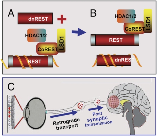
Fig. 6.
Schematic representation of the suppression of silencing of HSV DNA by dnREST. (A and B) Components of the repressor complex are assembled on the DNA and silence the DNA. dnREST bound to DNA precludes the binding of corepressors from binding to dnREST. In B, dnREST binds to DNA and precludes the assembly of the repressor complex. (C) Schematic representation of translocation of virus from the cornea to the brain. The virus is transported retrograde to the neuronal ganglion. Normally, the virus would establish latency and on reactivation be transported anterograde to the portal of entry (cornea). In the studies performed with the R112 recombinant virus, extensive replication and destruction of the ganglia could lead to postsynaptic transmission of the virus to the brain. In light of detection of virus in the brain of asymptomatic mice, it is conceivable that small amounts of virus “leak” into the brain on reactivation but that these do not cause symptomatic disease.

