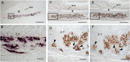
Fig. 3.
Histochemical characteristics of GPH producing cells in the adenohypophysis of mature hagfish. (A and B) Cellular expression of GPHα gene detected by in situ hybridization with labeled hagfish GPHα riboprobe. (C–F) Immunohistochemical detection of GPHα- and β-producing cells on adjacent pituitary sections detected with specific antisera: (C and D) anti-hagfish GPHα; (E and F) anti-hagfish GPHβ. Note that GPHα-producing cells are well in accordance with GPHβ-producing cells (arrowheads). B, D, and F show magnified views of regions shown by rectangles in A, C, and E, respectively. AH, adenohypophysis; CT, connective tissue; NH, neurohypophysis; III, third ventricle. (Scale bars: A, C, and E, 100 μm; B, D, and F, 20 μm.)

