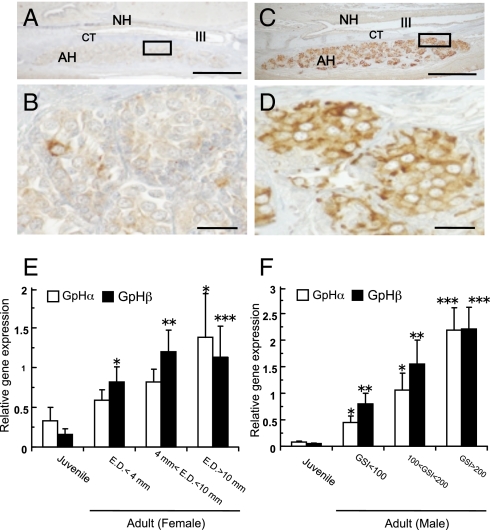
Fig. 4.
Correlation between pituitary GPH activities and gonadal development in hagfish. (A–D) Cellular activities of GPHβ cells in the hagfish pituitary. Note that intense immunoreactions are observed in mature female (C and D), but faint reactions presented in juvenile (A and B). AH, adenohypophysis; CT, connective tissue; NH, neurohypophysis; III, third ventricle. (Scale bars: A and C, 100 μm; B and D, 20 μm.) (E and F) Relative GPHα and GPHβ gene expressions in the pituitary of female (E) and male (F) hagfish. Open bars represent GPHα gene expressions and filled bars represent GPHβ gene expressions. The two GPH mRNA levels were normalized by β-actin mRNA levels. Relative values are expressed as mean ± SE (n = 8–18). Significant differences from the juvenile are indicated by *, P < 0.05; **, P < 0.01; and ***, P < 0.001. Note that two GPH transcripts in both sexes increase in accordance with the developmental stage of the gonad.

