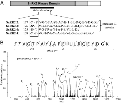
Fig. 1.
Increased phosphorylation of SnRK2 kinases following ABA treatment. (A) Peptides from the activation loop of the SnRK2 proteins have increased phosphorylation following ABA treatment (30 min). Asterisks indicate validated sites of phosphorylation. When the exact site of phosphorylation could not be determined from the MS2 spectra, all potentially modified residues are shown in italics. (B) MS2 spectra of the SnRK2.2 singly phosphorylated peptide following ABA treatment using ETD fragmentation. Fragment ions detected are shown with the phosphopeptide sequence and annotated in the spectrum. In this case, the phosphorylation site can be isolated only to the first two residues (as noted by italics) because of the lack of distinguishing fragment ions at the N terminus.

