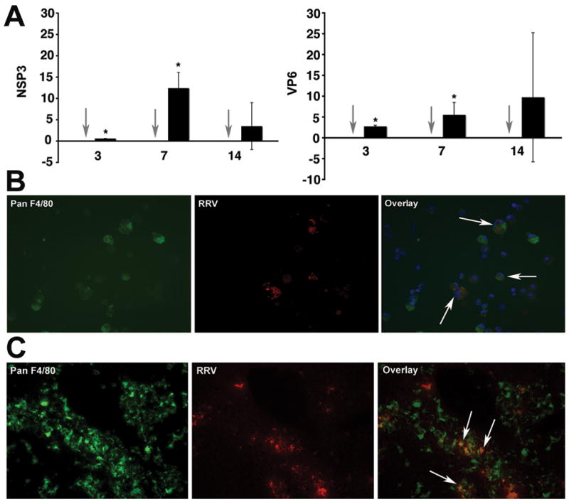
Figure 2. Detection of RRV in hepatic mononuclear cells and macrophages.

Panel A depicts the expression of mRNA encoding for the RRV proteins NSP3 and VP6 in hepatic mononuclear cells isolated from mice 7 days after RRV challenge. Grey arrows point to no detection of RRV in hepatic mononuclear cells of normal saline-injected controls; black bars=RRV injected mice; P<0.05; N=3–6 livers per time point and per experimental group. Panels B and C depict dual-immunofluorescence signals identifying RRV (red) in panF4/80+ cells (green) in purified hepatic mononuclear cells (panel B) or portal tracts (panel C) 7 days after RRV infection of newborn mice. Arrows point to double positive panF4/80+ and RRV+ cells.
