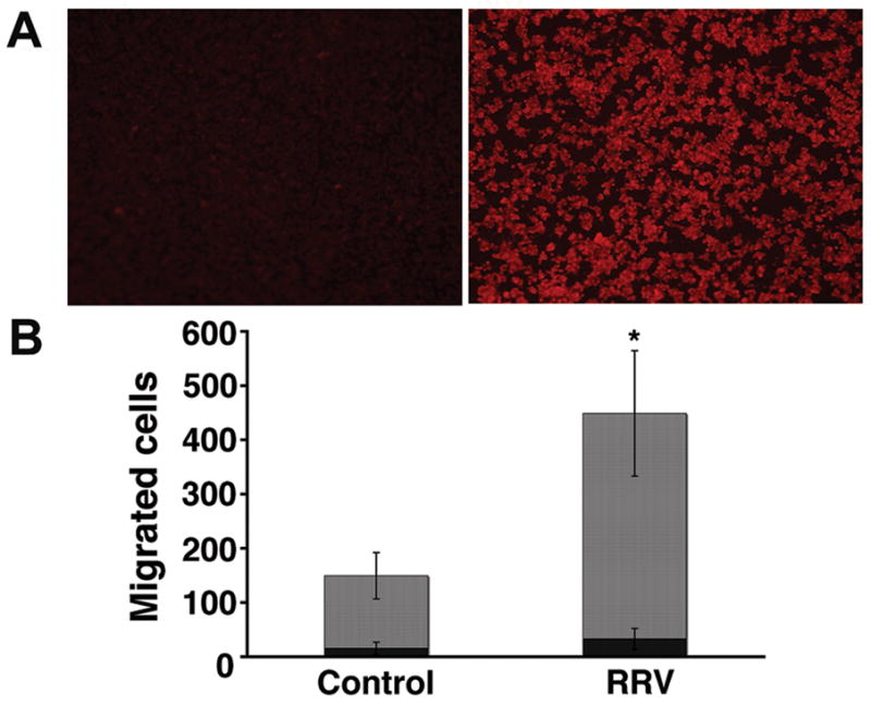
Figure 3. RRV infection of Raw 264.7 cells and induction of chemotaxis by conditioned media.

In panel A, immunofluorescence detects RRV (red signal) 24 hours after Raw 264.7 cells were exposed to RRV (right photograph); no signal is detected in cells not exposed to RRV (left photograph). Panel B depicts the numbers of neutrophils and lymphocytes that migrated to lower wells of chemotaxis chambers containing conditioned media from RRV-infected or control (RRV-naïve) Raw 264.7 cells after 45 minutes of culture. *P<0.05; N=3 chambers per group; grey bars=neutrophils; black bars=lymphocytes.
