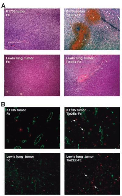
FIGURE 7.
Intratumoral injection of Tie2Ex in K1735 wild-type and Lewis lung tumors. Purified Tie2Ex-Fc or Fc in PBS was injected intratumorally into K1735 wild-type tumors or Lewis lung tumors. Tumors were excised 16 to 18 h after injection. A. H&E staining of tumor sections from K1735 wild-type tumors (top) or Lewis lung tumors (bottom) with Fc (left) or Tie2Ex-Fc (right). Total magnification, ×100. B. Frozen tumor sections from K1735 wild-type or Lewis lung tumors injected with Fc or Tie2Ex-Fc were stained with TUNEL (red) and CD31 (green) to identify apoptotic cells and vessels. Total magnification, ×100. Asterisks, regions of hemorrhage or cell death; arrows, apoptotic endothelial cells/vessels.

