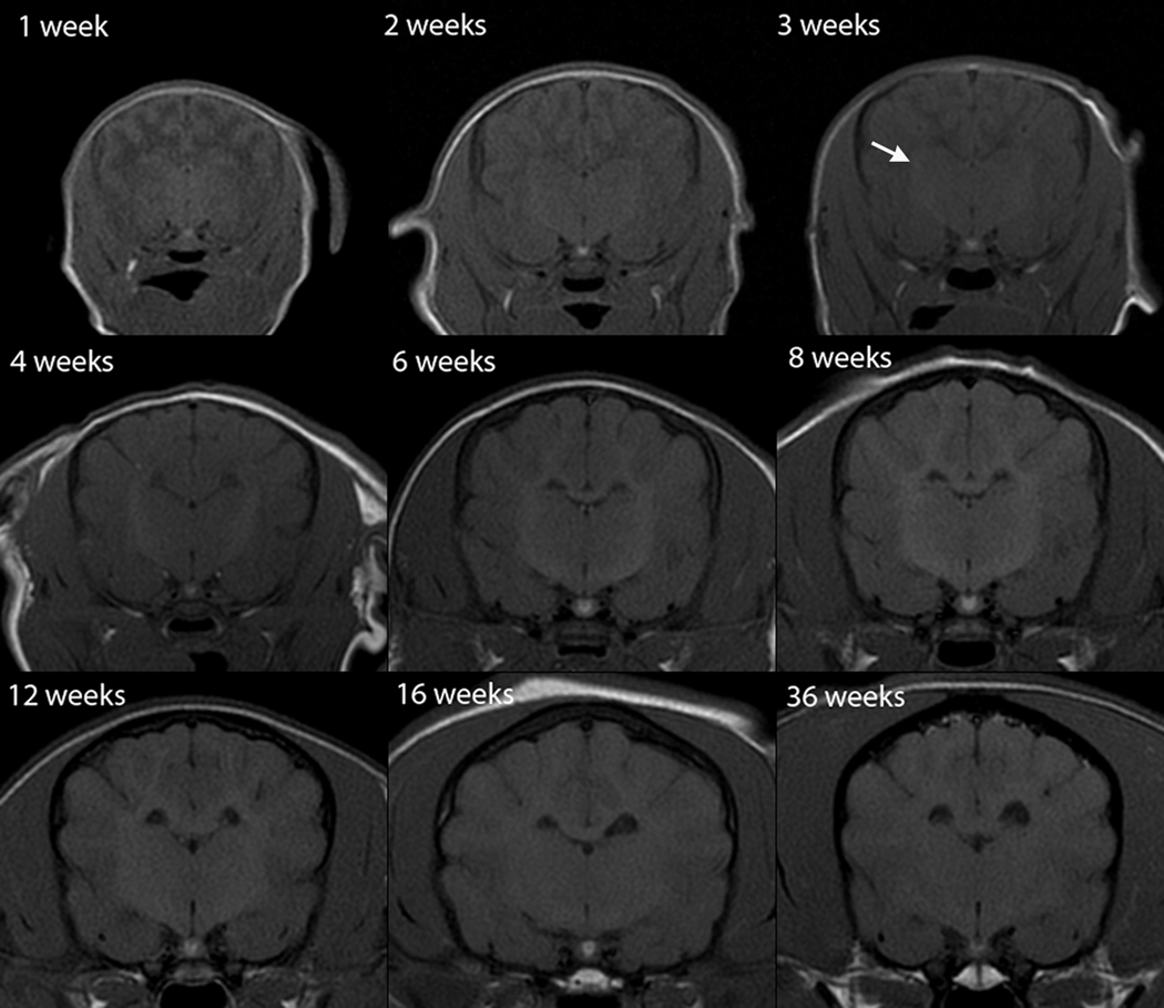
Fig. 6.
Transverse T1-weighted canine brain maturation progression. Image location is mid telencephalon, at the level of the interthalamic adhesion. The subcortical white matter is hypointense to gray matter during the juvenile phase (weeks 1–3), isointense during the transition phase (4 weeks) and exhibits hyperintensity during the maturing phase and into adulthood (6–36 weeks). The relative increase in white matter intensity is due to a combination of increasing lipid and decreasing water during myelination. The hyperintense internal capsule is first consistently identifiable at 3 weeks (arrow).

