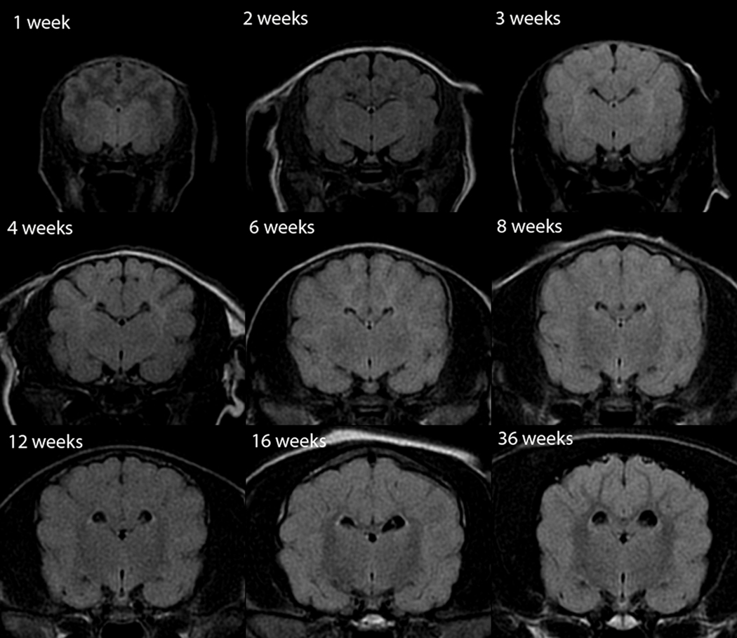
Fig. 8.
Transverse Fluid Attenuated Inversion Recovery (FLAIR) canine brain maturation progression. Image location is mid telencephalon, at the level of the interthalamic adhesion. The telencephalon juvenile phase is characterized by two sub-phases, each with an associated transition phase; this differs from the maturation appearance in T1W and T2W images. The subcortical white matter is hypointense to gray matter at weeks 1 and 2 with a transition to being isointense at 3 weeks. This is followed by hyperintensity at weeks 4 and 6 with a second isointensity phase at 8 weeks. From 12 weeks and older the subcortical white matter is characterized by a progressive hypointensity during the maturing phase. The initial period of subcortical white matter hypointensity is attributed to the large amount of free water resulting in a T1 relaxation time similar to CSF, with resultant nulling of the signal by the inversion pulse. Following the rapid initial decrease in free water during the first three weeks, there is not enough free water to result in suppression and the signal intensity follows the T2 relaxation changes as the water content continues to decrease during myelination.

