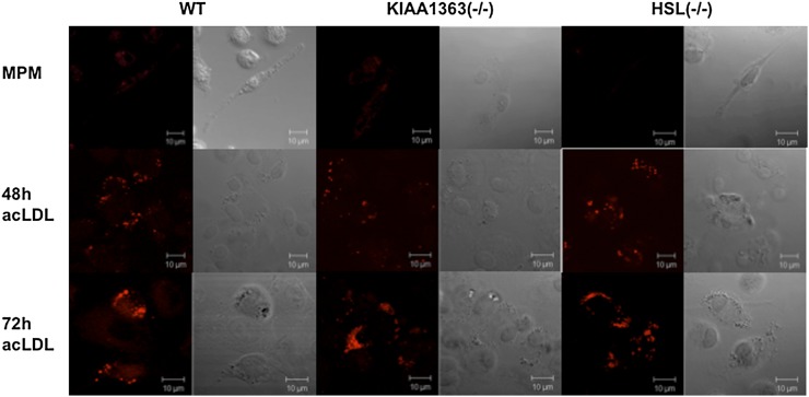
Fig. 8.
Foam cell formation of HSL−/− and KIAA1363−/− macrophages. Representative fluorescent microscopy after Nile red staining of HSL−/−, KIAA1363−/−, and WT macrophages and foam cells. Foam cell formation was achieved by loading macrophages with 100 µg acLDL for 48 h and 72 h. Images were taken on a Zeiss LSM 510 Meta microscope. HSL, hormone-sensitive lipase; MPM, mouse peritoneal macrophage; WT, wild-type.

