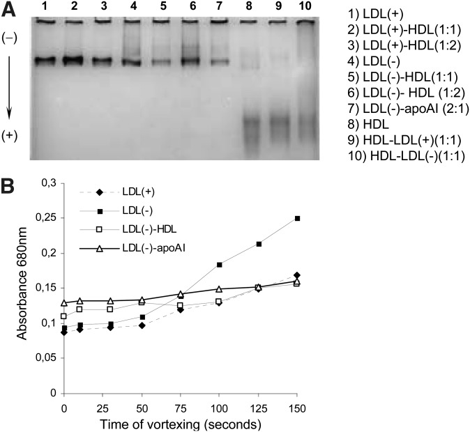
Fig. 5.
Basal aggregation and susceptibility to aggregation of LDL(+), LDL(–), and HDL in native state and after treatments. shows electrophoretic mobility in the experimental conditions in a GGE gel. Particles migrate in GGE gels according to their size and aggregates can be observed. The samples are LDL(+), LDL(–), and HDL in native state and after preincubation. The proportion of apoB:apoAI used in the preincubation is indicated: 1 = 0.5 g/L of apoB for LDL and 0.5 g/L of apoAI for HDL. The concentration used in the gel was 0.5 g/L of apoB or apoAI. shows a representative experiment of susceptibility to aggregation of LDL(+), LDL(–), and LDL(–) preincubated with HDL and apoAI (1:1). Absorbance at 680 nm was measured after increasing times of agitation by vortex.

