
Figure 5. The conformation of the 4c-4d loop of Semaphorins is central for Plexin recognition.
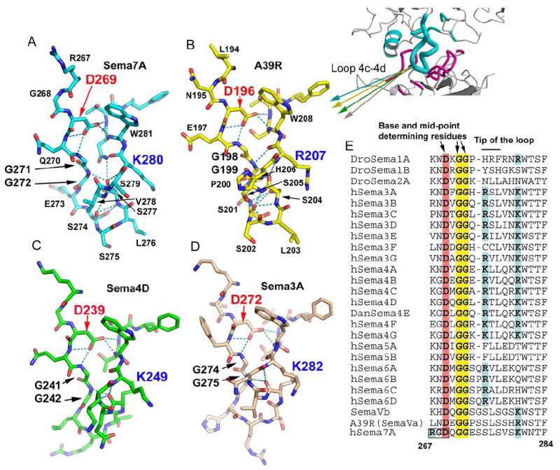
(A), (B), (C) and (D) Stick models of the isolated 4c-4d loops of Sema7A (cyan), A39R (yellow), Sema4D (green), and Sema3A (pink), showing its conserved conformation at the bases (top) and the mid-points of the loops, and variable conformation at the tips of the loops (bottom).
(E) Sequence comparison of the 4c-4d loop of Semaphorins, with the key conserved structural determinants highlighted.
See also Figure S5 for the RGD motif at the base of the Sema7A 4c-4d loop.