
Figure 6. Viral mimicry of Sema7A by A39R.
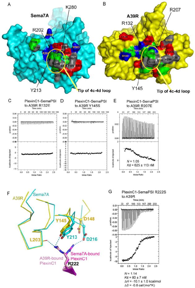
(A) and (B) The respective PlexinC1-binding surfaces of Sema7A (left, cyan) and A39R (right, yellow). The residues mapped at the interface are colored red for acidic residues, blue for basic residues, gray for polar, non-charged residues, and green for apolar residues.
(C), (D) and (E) Mutations of the A39R residues mapped to the Sema7A-PlexinC1 interface abolished or dramatically reduced A39R-PlexinC1 binding.
(F) The different conformation of PlexinC1 Arg222 in binding A39R and Sema7A.
(G) Calorimetric measurement of the binding between A39R and the Arg222Ser mutant of PlexinC1-SemaPSI.
A structural and sequence comparison of Sema7A and A39R is shown in Figure S3.