
Fig. 3. Immunohistology of frozen section of UHMWPE particle group and control group.
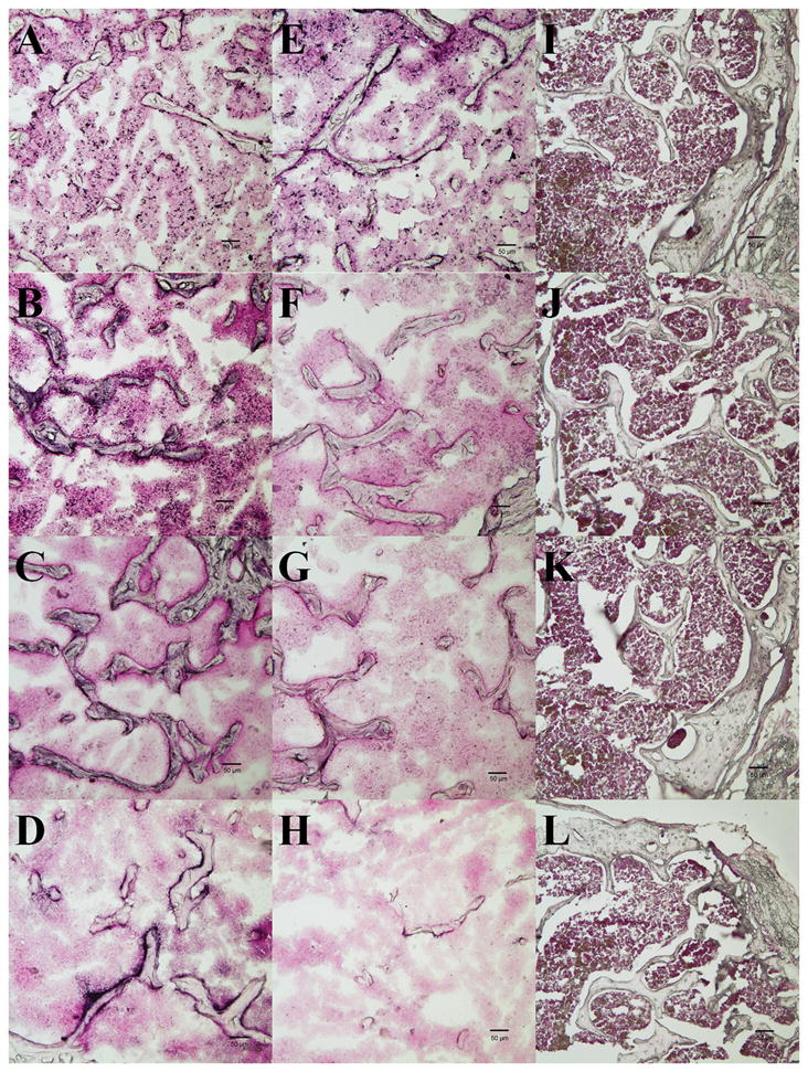
The first column (A–D) contains sections from UHMWPE particle injected femora; the 2nd column (E–H) contains sections from non-operated femora of UHMWPE particle groups; the 3rd column (I–L) contains sections from the saline injected femora. Sections in 1st row are stained with anti-CD68 antibody, 2nd – 4th rows are stained with anti-GFP, anti-osteocalcin, and anti-αVβ3 antibody, respectively. AP and its substrate BCIP/NBT were used for chromogenic reaction; Nuclear fast red was used for counter staining. Arrows in the panels C and D pointed the positive staining of anti-osteocalcin and anti-αVβ3. The scale bar length is 50 μm.