Figure 2.
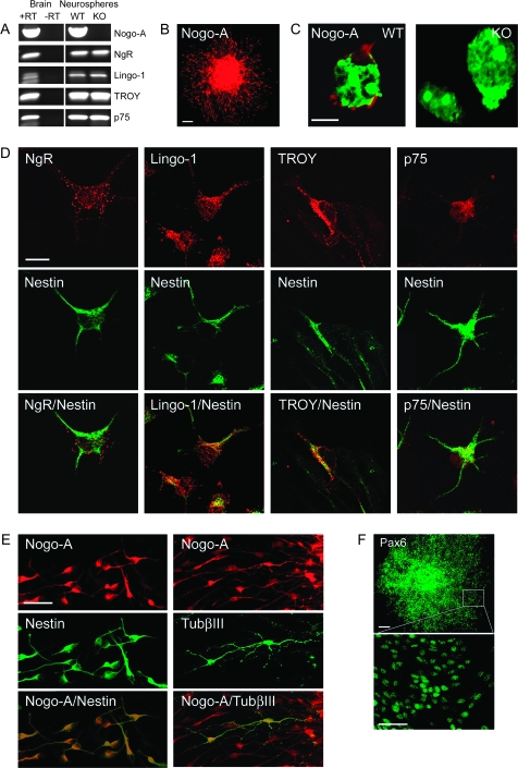
Nogo-A and the Nogo receptor components NgR, Lingo-1, TROY, and p75 are expressed by embryonic mouse forebrain–derived neurospheres. (A) Transcripts for Nogo-A, NgR, Lingo-1, TROY, and p75 are all detected by RT-PCR in E15.5 mouse forebrain–derived neurospheres (passage 5). Nogo-A is absent in Nogo-A KO mouse forebrain–derived neurospheres, but the receptor components persist. Total messenger RNA from neonatal mouse brain was used as a positive control. “+RT” and “−RT” indicate performance of reverse transcription with and without reverse transcriptase, respectively. (B) Nogo-A (red) immunoreactivity is found in precursor cells of a plated neurosphere cultured for 1 day without growth factors. Note the halo of migrating Nogo-A+ cells surrounding the sphere. (C) Localization of Nogo-A (red) on the cell surface of WT mouse neurosphere-derived cells. No immunostaining was observed on cells derived from Nogo-A KO mouse embryos. Nuclei are counterstained with green fluorescent Nissl stain. (D) Colocalization of NgR, Lingo-1, TROY, or p75 (red) and nestin (green) in migrating precursor cells emanating from plated neurospheres. (E) Colocalization of Nogo-A (red) and nestin (green) or TubβIII (green) in migrating precursor cells emanating from plated neurospheres. Approximately 1% of migrating cells are positive for TubβIII. (F) Expression of the radial cell glial marker Pax6 (green) in almost all neurosphere-derived cells. Scale bars: B = 100 μm; C = 10 μm; D = 10 μm; E = 50 μm; F = 100 μm, 50 μm.

