
Figure 1.
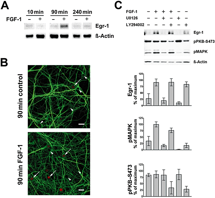
FGF-1 induces Egr-1 protein in primary hippocampal cell cultures. (A) The transcription factor Egr-1 was elevated in C3H mouse primary hippocampal neurons after 90 min of FGF-1 (10 ng·mL−1) treatment as judged by Egr-1 immunoreaction in the Western blot. Corresponding signals for β-actin are shown as loading control. (B) The number of Egr-1-positive nuclei (red) is elevated after application of FGF-1 for 90 min. Arrowheads indicate Egr-1-negative and arrows Egr-1-positive nuclei. An antibody against microtubule-associated protein 2 (MAP2, green) is used as a counter-stain (bars = 20 µm). (C) Upper panel: level of the transcription factor Egr-1, PKB and MAPK phosphorylation after 90 min of FGF-1-treatment (10 ng·mL–1) with or without the protein kinase inhibitors U0126 and LY294002 (both 10 µM) as shown in a representative Western blot. Lower panel: quantification of Western blot data for pMAPK, pPKB and Egr-1 normalized for β-actin (mean ± SD, n = 3).