
Figure 4.
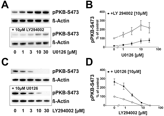
Effect of MEK-1/2 and PI3K inhibition on pPKB–S473 in HT22 cells. (A) Typical Western blot demonstrating that after 90 min, control samples displayed a basal level of pPKB–S473 which was elevated with increasing concentrations of U0126 (upper panel). Pre-incubation of 10 µM LY294002 (lower panel) led to a down-regulation of pPKB–S473. β-Actin immunoreaction served as loading control. (B) Analysis of β-actin-normalized pPKB signals showed that the maximum pPKB–S473 level was reached at 10 µM of the MEK-1/2 inhibitor. Pre-incubation of 10 µM LY294002 led to a down-regulation of pPKB–S473 in the absence of U0126 (P < 0.01). U0126 application led to an induction of pPKB–S473 with a maximum at 10 µM U0126 (P < 0.01) with no further induction at 30 µM U0126. Compared with the levels in the absence of LY294002, U0126-mediated induction of pPKB was generally suppressed in the presence of the PI3K inhibitor. Shown are the mean ± SD of two separate experiments with three repeats. (C) Typical Western blot showing that basal pPKB–S473 was reduced by increasing concentrations of LY294002 (upper panel). Pre-incubating 10 µM of the MEK-1/2 inhibitor U0126 induced pPKB–S473 levels which were reduced when rising concentrations of LY294002 were added. A full suppression of pPKB–S473 below the detection limit was seen at 30 µM PI3K inhibitor (10 µM U0126 + 30 µM LY294002). β-Actin immunoreaction served as loading control. (D) Statistical analysis of pPKB–S473 signals normalized to β-actin showing that basal pPKB–S473 was linearly reduced with increasing concentrations of LY294002. A reduction below detection limit was observed at 10 and 30 µM LY294002. U0126-mediated increase in pPKB–S473 was concentration-dependently reduced by the PI3K inhibitor. A full suppression was seen at 30 µM of LY294002. Shown are the mean ± SD of two separate experiments with three repeats.