
Figure 3.
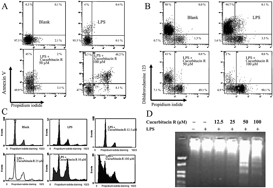
Cucurbitacin R induced apoptosis in RAW 264.7 cells in a concentration-dependent manner. Cells were treated for 24 h with cucurbitacin R (12.5–100 µM) after LPS stimulation. (A) Annexin V apoptosis assay. (B) Mitochondrial membrane potential assay. Cells were treated with cucurbitacin R at various concentrations for 18 h and apoptotic cells, apoptotic-necrotic cells, and necrotic cells were examined with annexin V or dihidrorodamine 123 and propidium iodide binding as described in the Methods section. (C) Plots were obtained with propidium iodide staining after cucurbitacin R treatment at different concentrations (12.5–100 µM). (D) DNA fragmentation assay in RAW 264.7 cells. Figures are representative of three experiments performed with similar results.