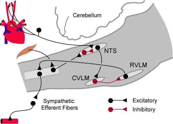
Figure 1.
A simplified schematic illustrating the brainstem baroreflex pathway. The baroreceptor afferent fibers, carrying blood pressure information, make excitatory synaptic contact with second-order neurons in the nucleus tractus solitarii (NTS), the first central site that receives and integrates the sensory inputs. The afferent fibers from skeletal muscle also project to the NTS through a poly-synapse pathway. These ascending fibers carrying information from the muscles make excitatory synaptic contact with the GABAergic interneurons in the NTS. The NTS output neurons convey signals from the baroreceptors and muscle afferents to neurons in the caudal ventral lateral medulla (CVLM) via excitatory glutamatergic synapses. The neuronal output of the CVLM provides the major inhibitory (GABAergic) inputs to the cardiovascular sympathetic neurons in the rostral ventral lateral medulla (RVLM), the major output neurons that regulate sympathetic nerve activity.

