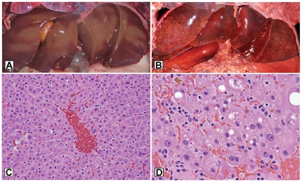
Figure 6. Gross and microscopic hepatic lesions of microcystin intoxication in sea otters, compared to control livers.
A.) Gross appearance of normal sea otter liver. B.) Swollen, hemorrhagic liver from a sea otter that died due to microcystin intoxication. C.) Microscopic view of normal sea otter liver, D.) Microscopic appearance of liver from an otter that died due to microcystin intoxication, demonstrating hepatocyte swelling, cytoplasmic vacuolation, necrosis or apoptosis and parenchymal hemorrhage. Small greenish-gold accumulations of bile are apparent at the upper left and center-right portions of the photomicrograph.

