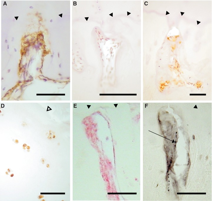
Fig. 4.
NGF expression in articular cartilage and bone. NGF-positive cells (brown) in vascular channels in the medial tibial plateau of patients with OA (A) and RA (C), but not in a non-arthritic control (B). (D) Chondrocytes displaying NGF immunoreactivity (brown) in superficial articular cartilage from a patient with OA (E and F). Co-localization within a vascular channel of NGF immunoreactivity (E, red) and a CGRP-immunoreactive nerve (F, black, arrow), demonstrated in serial tissue sections from a patient with OA. (A–D) DAB development with haematoxylin counterstain. (E) FastRed development. (F) Nickel-enhanced DAB development. Filled arrow heads: tidemark. Open arrow head: articular surface. Scale bar = 100 microns.

