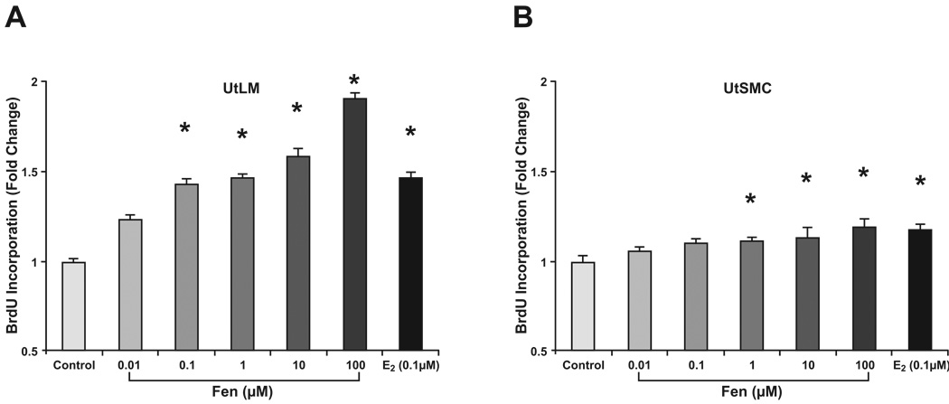
Figure 3. Cell proliferation assay with BrdU.
UtLM cells (A) and UtSMCs (B) exposed to Fen for 24 h at concentrations ranging from 0.01 µM to 100 µM. E2 at concentration of 0.1 µM was used as a positive control. Compared with proliferation in untreated controls, BrdU labeling in UtLM cells was increased at Fen concentrations in the 0.1 µM to 100 µM range. BrdU labeling in UtSMC cells was increased in the 1 µM to 100 µM range. Representative data were shown from two separate experiments. *p<0.01 compared with vehicle control cells.

