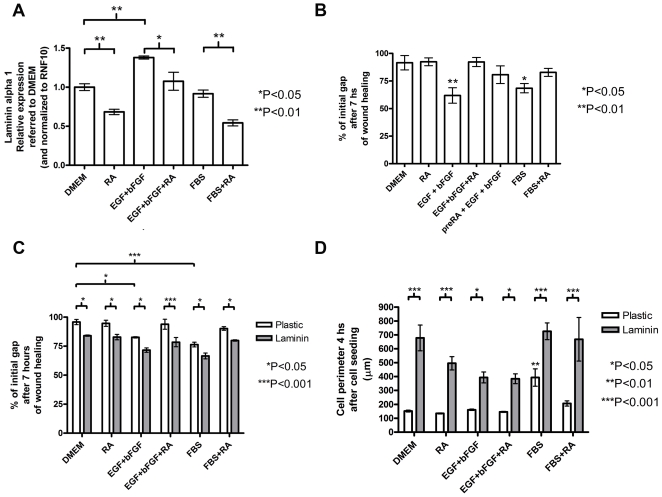
Figure 3. RA and EGF+bFGF treatments affect laminin expression which modulates cell migration and adhesion of hMSC.
A: QRT-PCR analysis of laminin alpha 1 expression in hMSC that were treated for 5 days with either DMEM, 0.5 µM RA in DMEM (RA), 20 ng/ml EGF+ 5 ng/ml bFGF (EGF+bFGF), 20 ng/ml EGF +5 ng/ml bFGF +0.5 µM RA (EGF+bFGF+RA), 10%FBS (FBS), or 0.5 µM RA in 10%FBS (FBS+RA). The error bars represent relative expression normalized to RNF10 expression and referred to the relative expression on DMEM as mean ± s.e.m. B: Wound healing experiments were performed on hMSC that were previously treated for 5 days as in A and also treated with 0.5 µM RA followed by EGF+bFGF at the time of the experiment (preRA+EGF+bFGF). The error bars represent % of the initial gap after 7 hours as mean ± s.e.m. C: Similar wound healing experiments as shown in B but hMSC were previously seeded over either laminin coated (20 µg/well) (laminin) or uncoated plates (plastic) and treated as in A. The error bars represent % of the initial gap after 7 hours as mean ± s.e.m. D: Cell spreading assay of hMSC that were treated for 5 days similarly as in A (see Materials and Methods). The error bars represent cell perimeter as mean ± s.e.m. All the treatments in A–D were performed in triplicates in two independent experiments with different hMSC donors and the significance of the results was assessed using one way ANOVA and Tukey's multiple comparison test (for A and B) or with two way ANOVA followed by Bonferroni post test (for C and D). *P<0.05; **P<0.01; ***P<0.001.

