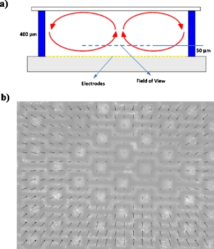
Figure 9.
(a) Schematic drawing of the side-view of the global flow observed in deep DEP chambers. (b) The velocity field measured inside the DEP chamber at a focal plane 50 μm above the electrode surface using the μ-PIV technique. The results are obtained in a 400 μm high DEP chamber using 1 μm fluorescent particles suspended in apple juice (0.225 S∕m) under 10 Vp.p. electric field at 10 MHz. The maximum measured velocity is 10 μm∕s.

