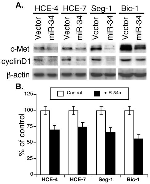
Figure 2.
miR-34a suppression of gene expression and proliferation of esophageal cancer cells. A, Western blotting. Esophageal squamous cell carcinoma lines HCE-4 and HCE-7 and adenocarcinoma cell lines Seg-1 and Bic-1 were grown in monolayer and transiently transfected with pRNA-u6.3/miR-34a vector or vector-only control and treated with G418 for 3 days. The total cellular protein was then extracted and subjected to Western blotting analysis of c-Met and cyclin D1 expression. B, Cell proliferation assay. Cells were grown in monolayer and transiently transfected with either pCMS/EGFP plus pRNA-u6.3/miR-34a vector or pCMS/EGFP plus pRNA-u6.3 vector. Cells were then fixed with 4% paraformaldehyde and permeabilized in Triton X-100. Afterward, cells were immunostained for Ki-67 expression, and approximately 200 cells were counted for positive GFP staining f (green) and positive or negative Ki-67 staining (red). The percentage of control of cell proliferation was then calculated.

